Figure Drawing - Individual Muscles - Neck
Số trang: 10
Loại file: pdf
Dung lượng: 1.09 MB
Lượt xem: 38
Lượt tải: 0
Xem trước 2 trang đầu tiên của tài liệu này:
Thông tin tài liệu:
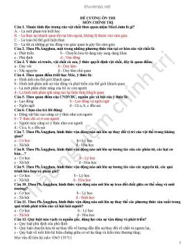
Short Neck Muscles The following three muscles are located on the back of the neck, just behind the skull: the obliquus capitis caudalis, the obliquus capitis cranialis, and the rectus capitis dorsalis major. They are covered by narrow and wide tendons and thin muscles, yet they help create the fullness on the back of the neck, determined in large part by the width of the atlas (the first neck vertebra) and the vertical projection of the axis (the second neck vertebra). Obliquus capitis caudalis (Large oblique muscle, Axoido-atloideus) • Origin: Entire side of the expanded upright spine of the...
Nội dung trích xuất từ tài liệu:
Figure Drawing - Individual Muscles - Neck42 INDIVIDUAL MUSCLES » NECK HORSE HORSE DOG TOP VIEW TOP VIEW DOG Short Neck Muscles Obliquus capitis cranialis (Small oblique muscle,The following three muscles are located on the back of the neck, just Atloido-occipitalis)behind the skull: the obliquus capitis caudalis, the obliquus capitis cra- • Origin: Front surface of the wing of the first neck vertebra (atlas).nialis, and the rectus capitis dorsalis major. They are covered by narrow • Insertion: Rear part of the skull.and wide tendons and thin muscles, yet they help create the fullness • Action: Both sides together extend the head.on the back of the neck, determined in large part by the width of the • Structure: This is a short muscle which fill the space between the skullatlas (the first neck vertebra) and the vertical projection of the axis (the and the first neck vertebra. It is directed forward, upward, and inward.second neck vertebra). Rectus capitis dorsalis major (Posterior straight muscle, Obliquus capitis caudalis (Large oblique muscle, Axoido-occipitalis) Axoido-atloideus) • Origin: Upper edge of the upright spine of the second neck vertebra.• Origin: Entire side of the expanded upright spine of the second neck • Insertion: Rear end of the skull near the midline.vertebra (axis). • Action: Extends the head.• Insertion: Rear surface of the expanded side projection, or wing, of the • Structure: This narrow muscle lies just to the side of, and partly under,first neck vertebra (atlas). the nuchal ligament of the neck in the horse and ox. In the dog and the• Action: Rotates the first neck vertebra (which pivots on the second neck feline, it lies against its fellow of the other side on the midline; thevertebra) to the side, thereby turning the head to the side. nuchal ligament begins at the rear of the second neck vertebra.• Structure: Largest of the group, this thick muscle is directed forwardand outward. Its rear portion is buried in muscle, but as it advances,it approaches the surface. INDIVIDUAL MUSCLES > NECK 43 HORSE HORSE Longissimus capitis and Longissimus atlantis end of the wing of the atlas by a strong, round tendon, which canHORSE become quite prominent on the surface. This tendon inserts in common• Origin: By tendinous fibers from the region of the sides of the first and with the splenius and the omotransversarius.second thoracic vertebrae, and by successive attachments from the The longissimus capitis and atlantis in the dog and feline do notupper sides of the seventh through the third neck vertebrae. affect surface form, but their tendons may be seen in the ox.• Insertion: Longissimus capitis: base of the skull behind the ear hole.Longissimus atlantis: lower end of the expanded side projection, or Several narrow or wide tendons and thin muscle pass over, or attachwing, of the first neck vertebra (atlas). onto the lower end of, the wing of the atlas. The deeper structures• Action: Muscles of both sides of the body: extend the head and neck. (splenius to the wing of the atlas, omotransversarius, longissimusOne side only: pulls the head and neck to that side, or rotates the atlas, atlantis, longissimus capitis, and semispinalis capitis) may showand therefore the head, to that side. through the more superficial structures (the wide, thin tendon and thin• Structure: The longissimus capitis and longissimus atlantis are two muscle of the brachiocephalicus and the wide, thin tendon of theelongated, parallel muscles, part of the longissimus system of the verte- splenius, both of which attach to the rear end of the skul ...
Tìm kiếm theo từ khóa liên quan:
Figure Drawing Individual Muscles Neck hội họa vẽ kỹ thuật vẽ nghệ thuật hội họa nghệ thuật vẽ văn hóa nghệ thuậtTài liệu có liên quan:
-
Bù sáng: Chụp tay không cài đặt
5 trang 234 0 0 -
Nghệ thuật sử dụng hiệu quả công cụ tài chính
3 trang 210 0 0 -
Khám phá những pho tượng độc, dị nhất Việt Nam
17 trang 204 1 0 -
Giáo trình Vẽ hình họa khối cơ bản và biến dạng - Trường Cao đẳng Lào Cai
36 trang 188 4 0 -
3 trang 161 0 0
-
14 trang 126 0 0
-
3 trang 119 0 0
-
The laws of black and white - Nguyên lý hội họa đen trắng: Phần 2
155 trang 117 2 0 -
3 trang 115 0 0
-
5 trang 113 0 0
-
1 trang 108 0 0
-
Thiết kế trình bày báo - 10 thủ thuật thiết kế báo in
5 trang 102 0 0 -
17 trang 93 0 0
-
Điêu khắc thời Trần (1225 – 1400)
17 trang 87 0 0 -
Giáo trình Đồ họa vi tính CorelDRAW nâng cao (Ngành: Hội họa) - Trường Cao đẳng Lào Cai
71 trang 83 1 0 -
Giáo trình Lịch sử Mỹ thuật (Ngành: Hội họa) - Trường Cao đẳng Lào Cai
77 trang 76 2 0 -
Sự khác nhau giữa bộ cảm biến hình ảnh CCD và CMOS trong máy ảnh kỹ thuật số
5 trang 74 0 0 -
7 lỗi cần tránh khi dùng email nơi công sở
4 trang 64 0 0 -
7 trang 62 1 0
-
4 trang 61 0 0